Innovative AI methods for medical imaging
The Biomedical Imaging Group Rotterdam develops AI methods to improve the analysis, reconstruction and quantification of medical images. Close collaboration with clinicians is one of the group’s hallmarks.
Since the invention of X-ray photography in 1895, the toolbox of medical imaging has grown impressively to include ultrasound, computed tomography and magnetic resonance imaging. The Biomedical Imaging Group Rotterdam (BIGR) aims to improve the efficiency and quality of state-of-the-art medical imaging by developing innovative AI methods. The group is part of the Erasmus MC, and chaired by associate professor Stefan Klein.
‘In essence, we develop AI-based software to help physicians interpret medical images’, says Klein. ‘We are a group of technical researchers. Whether it’s an image of the eye, the heart or the brain, ultimately, every image is a collection of pixels. So there’s a lot of overlap in the methods to analyse, reconstruct and quantify all these different image types.’
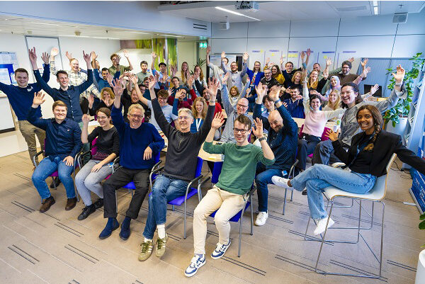
BIGR has over forty group members, including principal investigators, postdocs, PhD students, bachelor’s and master’s students and research software engineers. The group has a close link with the radiologists and other clinicians at the Erasmus MC. Klein: ‘We have a low-key cooperation, so we hear exactly what doctors need in practice. That helps us get clear on where to focus our AI models. For example, we might think that we should always be able to pinpoint exactly what kind of tumour is in an image. But sometimes doctors tell us it is not important in a specific case because it would not matter for treatment. However, in other cases, the precise differential diagnosis is crucial for clinical decision-making. The embedding within the Department of Radiology & Nuclear Medicine also gives us easier access to data and to scanning equipment, and we are closely collaborating with the MRI acquisition experts to improve image quality and reduce scan time.’
Klein has been working in the group since 2008. As one of the research highlights, he mentions the organisation of several Grand Challenges. Klein: ‘The first Grand Challenge I organised with my team in 2014 aimed to diagnose dementia early using MRI images. Fifteen international research teams participated in that challenge with 29 different methods. The accuracy turned out to be 63 percent, and the conclusion was that it should be a lot better. Still, that Grand Challenge had a lot of impact in determining exactly where the research field stood. In 2020, we organised a similar Grand Challenge for the diagnosis of osteoarthritis. Bringing people together in such challenges helps to push the field forward.’
Eye on the clinic
Luisa Sanchez is one of the two principal investigators of the eye image analysis research line of BIGR. She joined the group in 2018 after completing a PhD in computer vision. ‘What attracted me was the fact that it was a technical group in a hospital setting’, she says. ‘We are guided by what the clinic needs.’ Sanchez mainly focuses on image analysis of the retina. ‘In one of our projects, we are trying to find imaging biomarkers that can help clinicians track the progress of inherited retinal diseases. And as treatments for these diseases begin to emerge, we are also interested in seeing if these biomarkers can track the effect of treatment.’
Although the research group is large and works on a variety of medical applications, Sanchez says there are many advantages to functioning as a single research group. ‘Sometimes we want to link different organs and different technologies, like when we want to link brain biomarkers with retina biomarkers. Then, my collaborators and I can provide the expertise on retinal biomarkers and easily combine it with the brain biomarker expertise of the neuroimaging experts in BIGR. We encourage internal collaboration through group-wide activities, such as seminars and work groups on specific techniques that cut across different lines of research.’
Collaborating PhDs
Another way to best align BIGR’s technical-scientific research with clinical practice needs is to have a technical PhD student and a clinical PhD student working together on a project. ‘I see that yields many benefits in practice’, says Theo van Walsum, computer scientist and leader of the BIGR research line ‘Image guidance in interventions and therapy’.
‘Navigation is currently mainly used in the clinic in neurosurgery and orthopaedic surgery’, says Van Walsum about the research line. ‘There, you have the advantage that there hardly is any patient motion. Current navigation systems cannot handle changes in anatomy during the intervention. We are developing AI-based techniques that track instrument and patient motion. That way, physicians can use our technology in real-time to get their instruments to the right place in the best possible way. The aim is to integrate the 3D scan taken for diagnosis with the image taken live during the procedure. AI has given us a much more powerful toolbox that allows physicians to guide medical instruments faster and more effectively.’
Group passport – Biomedical Imaging Group Rotterdam (BIGR)
Research fields
Medical image analysis, machine learning, image reconstruction, quantitative imaging biomarkers, image-guided interventions
Institution
- Department of Radiology & Nuclear Medicine of Erasmus MC
Website
Published in I/O Magazine #1 2024
Text Bennie Mols
Images Ivar Pel
